CT CHEST EXAM TYPES
CT CHEST EXAM TYPES
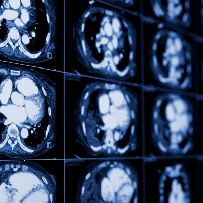
It is extremely important for the ordering clinician to understand the difference between a low-dose CT (LDCT) chest, a routine (diagnostic) CT chest, and a High Resolution CT Chest. We frequently encounter clinicians ordering a CT chest with contrast with thin slice imaging, since these are two different things all together I will explain the difference. A low-dose CT (LDCT), routine CT with contrast, and high-resolution CT (HRCT) of the chest serve different clinical purposes and vary in technical parameters.

LDCT is primarily used for lung cancer screening in asymptomatic, high-risk patients. It uses reduced radiation and no contrast, with thin slices (1–1.25 mm) to detect small lung nodules while minimizing risk from cumulative exposure. The thin slices overlap and essentially cover all the real estate of the lungs making this great for the radiologist to detect small nodules.
Alternatively, a routine CT chest with contrast is a diagnostic study used to evaluate known or suspected pathology such as infection, masses, vascular disease, or lymphadenopathy. It involves intravenous contrast material to enhance blood vessels and soft tissue, and typically uses higher radiation and standard slice thickness (~3–5 mm), with reconstructions available as needed. Because a slice is 5 mm thick, it can easily not detect a nodule that is 4 mm in size. Thus the clinical issue that sometimes the nodule is seen and sometimes it is not creating frustration for patients.
Meanwhile, high-resolution CT (HRCT) is a specialized protocol designed to evaluate diffuse or interstitial lung disease. It uses very thin slices (often 0.625 – 1.25 mm), high spatial resolution algorithms, and usually no contrast, focusing on lung parenchyma detail rather than soft tissue. Each slice is often only performed every 10 mm and thus all of the lung real estate is NOT covered. A 9 mm nodule will easily not be seen on this exam which as we all know is suspicious for malignancy. HRCT often includes images obtained in inspiration, expiration, and prone positioning to assess air trapping or dependent changes. The goal here is to evaluate the structures that are making up the lung parenchyma but NOT nodules.

